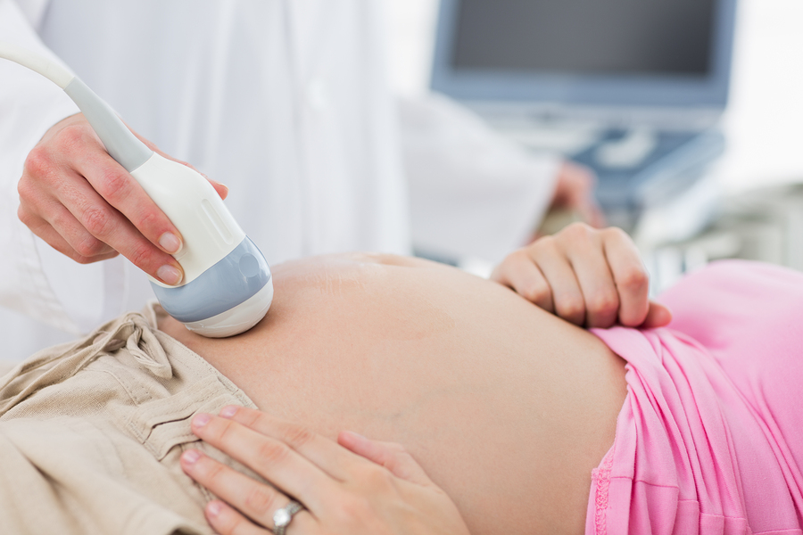
 Cleft lip and/or cleft palate can occur in a developing fetus for several reasons. One cause is genetic; the gene can be passed down from one or both parents. Drugs, viruses and toxins are some other causes of the birth defects. Sometimes the deformity is detected during pregnancy. High-tech ultrasonography allows doctors to view well-defined, two-dimensional images of a developing fetus. These high-resolution sonograms can reveal abnormalities of the face, including cleft lip. Although cleft palate is most often diagnosed after birth, in some cases ultra-precise three-dimensional ultrasonography and prenatal magnetic resonance imaging (MRI) may aid an in utero diagnosis.
By the fourth to sixth week of fetal development, the nose, lip, and part of the hard palate are formed. However, the facial features are not generally detectable with imaging equipment until 16-18 weeks. It is possible to diagnose a baby with cleft lip during a routine fetal check-up at approximately 18 weeks gestation. A second-trimester sonogram can show whether a baby has a normally shaped skull. Doctors look for signs that the skull has fused prematurely or if either side of the head is smaller than expected. Slight differences in facial structures are not always evident with prenatal ultrasonography. Many irregularities cannot be detected until after the baby is born.
If a second-trimester sonogram hints at a problem in the developing fetus, especially if there is a family history of a genetic disorder or syndrome chromosomal testing may be ordered. Pregnant women over the age of 35 are also routinely offered the option of chromosomal analysis. Amniocentesis and chorionic villus sampling can uncover inherited chromosomal disorders. With a very thin needle inserted into the abdomen, the doctor removes fetal cells from amniotic fluid or the tissue surrounding a newly developing embryo. The cells are sent to a lab where they grow and are then analyzed through a microscope. This process can uncover abnormalities related to a higher risk of craniofacial conditions.
While some clefts are diagnosed during pregnancy and others after a baby is born, a few more minor clefts are not as obvious and may not be diagnosed until later in life. Regardless of the cause or when they are diagnosed, the outlook for the treatment of clefts is a positive one. Although children with more severe cases will require more care and possibly multiple procedures, there are less invasive treatment options to restore a normal appearance and range of functions.
 Kayvon Haghighi, DDS, MD, FACS is licensed to practice both medicine and dentistry in the state of New Jersey. Dr. Haghighi’s unique combination of surgical training and experience in facial reconstruction enables him to analyze your condition from multiple points of view.